(MENAFN- PR Newswire)
Celebrating its golden anniversary, Nikon Small World honors a stunning photo of brain tumor cell structures, advancing our understanding of neurodegenerative diseases.
MELVILLE, N.Y., Oct. 17, 2024 /PRNewswire/ --
Nikon Instruments Inc. today announced the winners of the 50th annual Nikon Small World Photomicrography Competition, celebrating five decades of excellence in microscopy and digital imaging. This year's first place prize was awarded to Dr. Bruno Cisterna, with assistance from Dr. Eric Vitriol of Augusta University, for his groundbreaking image of differentiated mouse brain tumor cells, highlighting the actin cytoskeleton, microtubules, and nuclei. This image reveals how disruptions in the cell's cytoskeleton – the structural framework and "highways" known as microtubules – can lead to diseases like Alzheimer's and ALS.
Continue Reading
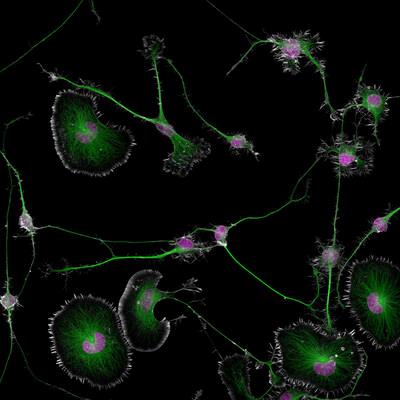
Differentiated mouse brain tumor cells (actin, microtubules, and nuclei)
Dr. Cisterna's research revealed that profilin 1 (PFN1), a protein crucial for building the cell's structure, plays a key role in maintaining the microtubule highways essential for cellular transport. When PFN1 or related processes are disrupted, these highways can malfunction, leading to cellular damage similar to what is observed in neurodegenerative diseases.
"One of the main problems with neurodegenerative diseases is that we don't fully understand what causes them," said Dr. Cisterna. "To develop effective treatments, we need to figure out the basics first. Our research is crucial for uncovering this knowledge and ultimately finding a cure. Differentiated cells could be used to study how mutations or toxic proteins that cause Alzheimer's or ALS alter neuronal morphology, as well as to screen potential drugs or gene therapies aimed at protecting neurons or restoring their function."
His patience and determination were crucial in capturing his image. "I spent about three months perfecting the staining process to ensure clear visibility of the cells. After allowing five days for the cells to differentiate, I had to find the right field of view where the differentiated and non-differentiated cells interacted. This took about three hours of precise observation under the microscope to capture the right moment, involving many attempts and countless hours of work to get it just right."
The hard work behind this discovery underscores its significance, bringing researchers closer to answers that could potentially transform millions of lives.
"After three years of research, we finally published our findings four months ago in the Journal of Cell Biology , and there's still more work to be done," said Dr. Cisterna. "I'm deeply passionate about scientific imaging; I've been following the Nikon Small World contest for about 15 years. It's an incredible contest that highlights the beauty of photomicrography but also inspires continued exploration and innovation in the field."
Eric Flem, Senior Manager, CRM and Communications at Nikon Instruments, shares a similar perspective on the competition. "At 50 years, Nikon Small World is more than just an imaging competition – it's become a gallery that pays tribute to the extraordinary individuals who make it possible. They are the driving force behind this event, masterfully blending science and art to reveal the wonders of the microscopic world and what we can learn from it to the public." He went on to add, "Sometimes, we overlook the tiny details of the world around us. Nikon Small World serves as a reminder to pause, appreciate the power and beauty of the little things, and to cultivate a deeper curiosity to explore and question."
Second place was awarded to Dr. Marcel Clemens
for his image of an electrical arc between a pin and a wire, produced by applying a potential difference of 10,000 volts.
Third place
was awarded to Chris Romaine for his image of a cannabis plant leaf. The bulbous structures are trichomes, or hair-like plant appendages, and the bubbles inside are cannabinoid vesicles, fluid-filled, blister-like structures.
In total, Nikon Small World recognized 87 photos out of thousands of entries from scientists and artists across the globe.
The 2024 judging panel included:
Adrian Coakley, Director of Photography at National Geographic Books
Michelle S. Itano, Ph.D. , Assistant Professor of Cell Biology and Physiology and Director of the Neuroscience Microscopy Core at the University of North Carolina at Chapel Hill
Emily Petersen , Photography Managing Editor at Science Magazine
Clare Waterman, Ph.D., Cell Biologist and Member of the National Academy of Sciences
Jennifer C. Waters, Ph.D., Director of the Core for Imaging Technology & Education at Harvard Medical School
Samantha Yammine, Ph.D., Neuroscientist and Science Communicator
For additional information, please visit , or follow the conversation on Facebook , Twitter @NikonSmallWorld and Instagram @NikonInstruments .
NIKON SMALL WORLD WINNERS
1st Place
Dr. Bruno Cisterna & Dr. Eric Vitriol
Medical College of Georgia at Augusta University
Department of Neuroscience & Regenerative Medicine
Augusta, Georgia, USA
Differentiated mouse brain tumor cells (actin, microtubules, and nuclei)
Super-Resolution
100X (Objective Lens Magnification)
2nd Place
Dr. Marcel Clemens
Verona, Veneto, Italy
Electrical arc between a pin and a wire
Image stacking for the pin and wire combined with long exposure for the electrical arcs
10X (Objective Lens Magnification)
3rd Place
Chris Romaine
Kandid Kush
Port Townsend, Washington, USA
Leaf of a cannabis plant. The bulbous glands are trichomes. The bubbles inside are cannabinoid vesicles.
Image Stacking
20X (Objective Lens Magnification)
4th Place
Dr. Amy Engevik
Medical University of South Carolina
Department of Regenerative Medicine & Cell Biology
Charleston, South Carolina, USA
Section of a small intestine of a mouse
Fluorescence
10X (Objective Lens Magnification)
5th Place
Thomas Barlow & Connor Gibbons
Columbia University
Department of Neurobiology and Behavior
New York, New York, USA
Cluster of octopus (Octopus hummelincki) eggs
Darkfield, Stereomicroscopy, Focus Stacking
3X (Objective Lens Magnification)
6th Place
Henri Koskinen
Helsinki University
Helsinki, Uudenmaan lääni, Finland
Slime mold (Cribraria cancellata)
Image Stacking, Polarized Light, Reflected Light
10X (Objective Lens Magnification)
7th Place
Gerhard Vlcek
Maria Enzersdorf, Austria
Cross section of European beach grass (Ammophila arenaria) leaf
Brightfield, Image Stacking
10X (Objective Lens Magnification)
8th Place
Stephanie Huang
Victoria University of Wellington
School of Biological Sciences; School of Psychology
Wellington, New Zealand
A neuron densely covered in dendritic spines from the striatum of an adult rat brain
Confocal, Deconvolution, Image Stacking
60X (Objective Lens Magnification)
9th Place
John-Oliver Dum
Medienbunker Produktion
Bendorf, Rheinland Pfalz, Germany
Pollen in a garden spider (Araneus) web
Image Stacking
20X (Objective Lens Magnification)
10th Place
Jan Martinek
Charles University
Department of Experimental Plant Biology
Prague, Czech Republic
Spores of black truffle (Tuber melanosporum)
Confocal
63X (Objective Lens Magnification)
11th Place
Dr. Ferenc Halmos
Bánd, Veszprém, Hungary
Slime mold on a rotten twig with water droplets
Image Stacking
0.7X - 4.5X (Objective Lens Magnification)
12th Place
Daniel Knop
Oberzent-Airlenbach, Hessen, Germany
Wing scales of a butterfly (Papilio ulysses) on a medical syringe needle
Image Stacking
20X (Objective Lens Magnification)
13th Place
Paweł Błachowicz
Bedlno, Świętokrzyskie, Poland
Eyes of green crab spider (Diaea dorsata)
Image Stacking, Reflected Light
20X (Objective Lens Magnification)
14th Place
Marek Miś
Marek Miś Photography
Suwalki, Podlaskie, Poland
Recrystallized mixture of hydroquinone and myoinositol
Polarized Light
10X (Objective Lens Magnification)
15th Place
Sébastien Malo
Saint Lys, Haute-Garonne, France
Isolated scales on Madagascan sunset moth wing (Chrysiridia ripheus)
Darkfield, Image Stacking, Reflected Light
40X (Objective Lens Magnification)
16th Place
Marek Miś
Marek Miś Photography
Suwalki, Podlaskie, Poland
Two water fleas (Daphnia sp.) with embryos (left) and eggs (right)
Darkfield, Polarized Light
10X (Objective Lens Magnification)
17th Place
Dr. Frantisek Bednar
Svosov, Zilinsky, Slovak Republic
Stonewort algae (Chara virgata) reproductive organs - oogonia (female organs) and antheridia (male organs)
Darkfield
4X (Objective Lens Magnification)
18th Place
Alison Pollack
San Anselmo, California, USA
An insect egg parasitized by a wasp
Image Stacking, Reflected Light
10X (Objective Lens Magnification)
19th Place
Alison Pollack
San Anselmo, California, USA
Seed of a Silene plant
Image Stacking, Reflected Light
10X (Objective Lens Magnification)
20th Place
Dr. Bruno Cisterna & Dr. Eric Vitriol
Medical College of Georgia at Augusta University
Department of Neuroscience & Regenerative Medicine
Augusta, Georgia, USA
Early stage of mouse neuroblastoma
cell differentiation (actin, microtubules, and mitochondria)
Super-Resolution
100X (Objective Lens Magnification)
HM
Christopher Algar
Hounslow, Middlesex, United Kingdom
Brine shrimp
Darkfield, Image Stacking, Polarized Light
4X (Objective Lens Magnification)
HM
Dr. Kseniia Bondarenko
University of Edinburgh
Institute for Immunology and Infection Research
Edinburgh, MidLothian, United Kingdom
Acute-stage parasites of Toxoplasma gondii in a human skin cell
Expansion Microscopy, Confocal, Deconvolution
100X (Objective Lens Magnification)
HM
Dr. Anja de Lange
University of Cape Town
Neuroscience Institute & Department of Human Biology
Cape Town, Western Cape, South Africa
Astrocytes surrounding a blood vessel in a thin slice of human brain
Confocal
40X (Objective Lens Magnification)
HM
Dr. Amy Engevik
Medical University of South Carolina
Department of Regenerative Medicine & Cell Biology
Charleston, South Carolina, USA
Intestinal villi
Fluorescence
20X (Objective Lens Magnification)
HM
Daniel Evrard
Aywaille, Liege, Belgium
Vinyl player needle on scratched vinyl disk
Image Stacking, Polarized Light
20X (Objective Lens Magnification)
HM
Randy Fullbright
Fullbright Studio
Vernal, Utah, USA
Agatized dinosaur bone
Image Stacking
10X (Objective Lens Magnification)
HM
Dr. David Maitland
St. Andrews, Fife, United Kingdom
Transverse section of rachis (stem) of bracken fern (Pteridium aquilinum)
Differential Interference Contrast (DIC)
5X (Objective Lens Magnification)
HM
Angus Rae
Australian National University
Centre for Advanced Microscopy
MacGregor, Australian Capital Territory, Australia
Autofluorescence in the face of a little two-spotted ladybird (Diomus notescens) Fluorescence lifetime imaging microscopy (FLIM)
20X (Objective Lens Magnification)
HM
Dr. Igor Robert Siwanowicz
Howard Hughes Medical Institute (HHMI), Janelia Research Campus
Ashburn, Virginia, USA
Antenna of a mole crab
Confocal
10X (Objective Lens Magnification)
HM
Jochen Stern
Mannheim, Baden-Wuerttemberg, Germany
Golden bug eggs on a sage leaf
Image Stacking
20X (Objective Lens Magnification)
HM
Dr. Bruce Douglas Taubert
Glendale, Arizona, USA
Ocelli between the compound eyes of a yellow jacket
Reflected Light
10X (Objective Lens Magnification)
HM
Kevin Terretaz
CRBM-CNRS
Montpellier, Hérault, France
Mosquito cells in culture with fluorescent markers for DNA and microtubules Confocal
63X (Objective Lens Magnification)
IoD
Dr. Sherif Abdallah Ahmed
Tanta University, Faculty of Science
Department of Zoology
Tanta, Egypt, Arab Republic
Anterior section of palm weevil
Image Stacking
4X (Objective Lens Magnification)
IoD
Anne Patricia Algar
Hounslow, Middlesex, United Kingdom
Mosquito larva
Darkfield, Image Stacking, Polarized Light
4X (Objective Lens Magnification)
IoD
Dr. Florian Alonso
University of Bordeaux
BioTis-INSERM U1026
Pessac, Gironde, France
Mouse aortic endothelium stained for beta-catenin (green), laminin (purple), smooth muscle actin (red), and Hoechst (cyan)
Confocal
63X (Objective Lens Magnification)
IoD
Didier Barbet
Club Français de Microscopie
Bailly, France
Fracture surface of mica (mineral)
Differential Interference Contrast (DIC)
10X (Objective Lens Magnification)
IoD
Timothy Boomer
WildMacro
Vacaville, California, USA
Slime mold (Prototrichia metallica)
Image Stacking
10X (Objective Lens Magnification)
IoD
Zhang Chao
National Astronomical Observatories, Chinese Academy of Sciences
Beijing, China
Beach sand
Reflected Light
10X (Objective Lens Magnification)
IoD
Joshua Coogler
Dallas, North Carolina, USA
Moss sporophyte with spores (green)
Image Stacking
10X (Objective Lens Magnification)
IoD
Nikky Corthout & Miranda Dyson
VIB (Flanders Institute of Biotechnology)
Center for Brain and Disease Research
Leuven, Vlaams-Brabant, Belgium
Fruit fly (Drosophila) brain vasculature
Confocal, Fluorescence, Image Stacking, Super-Resolution
25X (Objective Lens Magnification)
IoD
Nadia Efimova
Amicus Therapeutics
Philadelphia, Pennsylvania, USA
Dandelion pappus
Confocal, Fluorescence
20X (Objective Lens Magnification)
IoD
Dr. Laurent Formery & Dr. Nathaniel Clarke
Stanford University
Department of Molecular and Cell Biology
Pacific Grove, California, USA
Nervous system of a young sea star
Confocal, Fluorescence
10X (Objective Lens Magnification)
IoD
Karl Gaff
Dublin, Ireland
Larva of a midge fly (Chironomidae)
Darkfield, Image Stacking, Polarized Light
20X (Objective Lens Magnification)
IoD
Dr. Nick Gatford
University of Oxford
Nuffield Department of Clinical Neurosciences (NDCN)
Oxford, Oxfordshire, United Kingdom
A network of dopaminergic neurons generated from human stem cells
Super-Resolution
63X (Objective Lens Magnification)
IoD
Dr. Saikat Ghosh
National Institutes of Health
NICHD
Bethesda, Maryland, USA
Human neurons
Confocal
40X (Objective Lens Magnification)
IoD
Gerd A. Günther
Düsseldorf, Germany
Cross section of a beach grass (Ammophila arenaria) leaf
Fluorescence
20X (Objective Lens Magnification)
IoD
Anna-Mari Elisabeth Haapanen-Saaristo
University of Turku
Turku Bioscience Centre / Cell Imaging & Cytometry Core and Zebrafish Core RAISIO, Varsinais-Suomi, Finland
Nutrient storage cells in a tardigrade
Confocal
25X (Objective Lens Magnification)
IoD
Dr. Martin Hein
Lions Eye Institute
Physiology and Pharmacology laboratory
Nedlands, Western Australia, Australia
Abnormal blood vessel formation in a human retina with severe diabetic retinopathy Confocal
20X (Objective Lens Magnification)
IoD
Wen Jie Ji
Yin Works
The Bureau of Microworld Exploration
Beijing, China
Integrated circuit chip
Reflected Light
10X (Objective Lens Magnification)
IoD
Ted Kinsman
Rochester Institute of Technology
Photosciences Department
Rochester, New York, USA
A common house cat claw
Polarized Light
10X (Objective Lens Magnification)
IoD
Daniel Knop
Oberzent-Airlenbach, Hessen, Germany
Opening of a hibiscus flower (Hibiscus moscheutos) exposing the pollen in four stages, each ten minutes apart
Image Stacking
20X (Objective Lens Magnification)
IoD
Daniel Knop
Oberzent-Airlenbach, Hessen, Germany
Dorsal part of cuckoo wasp (Hedychrum gerstaeckeri) abdomen
Image Stacking
20X (Objective Lens Magnification)
IoD
Dr. Håkan Kvarnström
Bromma, Sweden
Peacock plume feather
Epi-Illumination, Image Stacking
4X (Objective Lens Magnification)
IoD
Dr. Ewa Langner
Washington University in St Louis
Department of Medicine - Renal Division, Mahjoub Lab
St Louis, Missouri, USA
Mouse embryonic kidney showing interstitial fibroblasts (yellow), tubular epithelium (cyan), and nuclei (magenta)
Confocal, Fluorescence, Image Stacking
40X (Objective Lens Magnification)
IoD
Dr. Amir Maqbool
Lovely Professional University
Department of Zoology
Srinagar, Jammu and Kashmir, India
Small fly killed by "zombie fly" fungus (Entomophthora muscae)
Image Stacking
2X (Objective Lens Magnification)
IoD
Dr. Robert Markus
University of Nottingham
School of Life Sciences, Super Resolution Microscopy
Nottingham, Nottinghamshire, United Kingdom
Dandelion (Traxacum officinale) cross section showing curved stigma with pollen
Confocal
10X (Objective Lens Magnification)
IoD
Dr. Robert Markus, Dr. Zeeshan Mohammad, Dr. Sarah Pashley & Dr. Rita Tewari University of Nottingham
School of Life Sciences, Super Resolution Microscopy
Nottingham, Nottinghamshire, United Kingdom
Malaria parasites and mouse blood cells - tubulin (green), all proteins (purple), DNA (red)
Confocal
63X (Objective Lens Magnification)
IoD
Jan Martinek
Charles University
Department of Experimental Plant Biology
Prague, Czech Republic
Spores of a black Bagnoli truffle (Tuber mesentericum)
Confocal
63X (Objective Lens Magnification)
IoD
Dr. Guillermo Moya
Johns Hopkins University
Department of Biology
Baltimore, Maryland, USA
Neuronal axons connecting to the muscles of the iris and the cornea
Confocal, Fluorescence
10X (Objective Lens Magnification)
IoD
Jacek Myslowski
Wloclawek, Kujawko-Pomorskie, Poland
Water mite (Arrenurus)
Fluorescence, Image Stacking
6.3X (Objective Lens Magnification)
IoD
Aryah Nagarajan
Falmouth University
Institute of Photography
Penryn, Cornwall, United Kingdom
Spores releasing from the sori of a Polypody fern (Polypodium vulgare)
Reflected Light, Transmitted Light, Focus Stacking
10X (Objective Lens Magnification)
IoD
Thomas Neumann
Tübingen, Baden-Württemberg, Germany
Ink dot on Japanese washi paper
Brightfield, Image Stacking
20X (Objective Lens Magnification)
IoD
Satu Paavonsalo & Dr. Sinem Karaman
University of Helsinki
Individualized Drug Therapy Research Program, Faculty of Medicine, Helsinki, Finland Helsinki, Finland
Blood vessels (color gradient) and endothelial cell nuclei (white) in the intestinal villi of a mouse
Confocal
10X (Objective Lens Magnification)
IoD
Uwe Lange
Hannover, Niedersachsen, Germany
Pollen on the compound eyes of a fly
Image Stacking
60X (Objective Lens Magnification)
IoD
Dr. Marko Pende
MDI Biological Laboratory
Murawala Lab
Bar Harbor, Maine, USA
Ladybug (Coccinellidae) on a clover (Trifolium repens)
Confocal, Fluorescence, Image Stacking
4X (Objective Lens Magnification)
IoD
Dr. Felice Placenti
FP Nature and Landscape Photography
Siracusa, Sicilia, Italy
Potato tuber sprout
Image Stacking, Reflected Light
1X (Objective Lens Magnification)
IoD
Alison Pollack
San Anselmo, California, USA
Slime mold (Lamproderma arcyrioides)
Image Stacking, Reflected Light
10X (Objective Lens Magnification)
IoD
Dr. Gonzalo Quiroga Artigas
CRBM-CNRS
Montpellier, Herault, France
Tardigrade (Hypsibius exemplaris)
Confocal
40X (Objective Lens Magnification)
IoD
Chris Romaine
Kandid Kush
Port Townsend, Washington, USA
Bract (part of the plants reproductive structures) of a cannabis plant. The bulbous glands are trichomes.
Image Stacking
20X (Objective Lens Magnification)
IoD
Dr. Adolfo Ruiz De Segovia
PARTICULAR
Madrid, Spain
Plant root
Fiber Optic
2X (Objective Lens Magnification)
IoD
Yurim Seo, Dr. Mark Looney & Dr. Simon Cleary
University of California, San Francisco
Pulmonary, Critical Care, Allergy and Sleep Medicine
San Francisco, California, USA
Lymphatic vasculature (cyan) and vessels (red) of a mouse lung
Confocal
4X (Objective Lens Magnification)
IoD
Dr. Leo Serra
University of Cambridge
Sainsbury Laboratory
Cambridge, Cambridgeshire, United Kingdom
Leaves arising from thale cress (Arabidopsis thaliana) meristem
Confocal
20X (Objective Lens Magnification)
IoD
Dr. Igor Robert Siwanowicz
Howard Hughes Medical Institute (HHMI), Janelia Research Campus
Ashburn, Virginia, USA
Aster anther cross section with pollen grains (green)
Confocal
40X (Objective Lens Magnification)
IoD
Dr. Igor Robert Siwanowicz
Howard Hughes Medical Institute (HHMI), Janelia Research Campus
Ashburn, Virginia, USA
Floret of a common chicory with pollen grains (spiky balls)
Confocal
40x (Objective Lens Magnification)
IoD
Luna Šošo Zdravković, Michael Surala & Christian Madry
Charité Universitätsmedizin Berlin
Institute of Neurophysiology
Berlin, Germany
Pyramidal neuron in mouse hippocampus
Confocal, Fluorescence, Image Stacking
30X (Objective Lens Magnification)
IoD
Dr. Bruce Douglas Taubert
Glendale, Arizona, USA
Mid-tibial tuft on a male orchid bee, used to attract mates
Reflected Light
10X (Objective Lens Magnification)
IoD
Maxime Teixeira
Laval University
Department of Molecular Medicine
Québec, Canada
Cultured monkey kidney cells labeled for tubulin (blue) and actin (orange) showing pathological accumulation of alpha-syn aggregates (red)
Confocal, Deconvolution, Image Stacking, Super-Resolution
100X (Objective Lens Magnification)
IoD
Dr. Theo Theune
Oost-Souburg, Zeeland, Netherlands
Abdominal skin of a tick that engorged with blood
Image Stacking
50X (Objective Lens Magnification)
IoD
Dr. Grigorii Timin & Dr. Michel Milinkovitch
University of Geneva
Department of Genetics and Evolution
Geneva, Switzerland
Skin scales of a snake embryo stained with Fast Green dye
Confocal
63X (Objective Lens Magnification)
IoD
Steven A. Valley
Oregon Department of Agriculture (ODA)
Entomology Lab
Albany, Oregon, USA
Immature male damselfly (Calopteryx aequabilis)
Image Stacking, Reflected Light
5X (Objective Lens Magnification)
IoD
Dr. Bruno Vellutini
Max Planck Institute of Molecular Cell Biology and Genetics
Dresden, Saxony, Germany
Gene expression patterns in a drain fly embryo (Clogmia albipunctata) with an open eggshell
Confocal
20X (Objective Lens Magnification)
IoD
Susannah Waxman & Dr. Ian Sigal
University of Pittsburgh
Department of Ophthalmology
Pittsburgh, Pennsylvania, USA
Optic nerve head collagen of a pig
Image Stacking, Multiphoton
25X (Objective Lens Magnification)
IoD
Shao Yang
Beijing Miteyide Culture Co., Ltd.
Beijing, China
Fiber of nylon stockings
Polarized Light
5X (Objective Lens Magnification)
IoD
Chew Yen Fook
Woodend, Waimakiriri, New Zealand
Graffiti from Berlin Wall stone section
Image Stacking, Top Illumination
10X (Objective Lens Magnification)
IoD
Chew Yen Fook
Woodend, Waimakiriri, New Zealand
Water mite (Hydrachna sp.)
Darkfield, Image Stacking, Polarized Light
20X (Objective Lens Magnification)
IoD
Elkhan Yusifov & Dr. Martina Schaettin
University of Zurich
Department of Molecular Life Sciences
Zurich, Switzerland
Developing nervous system in the eye of a 7-day-old chick embryo
Stereomicroscopy
10X (Objective Lens Magnification)
IoD
Ou Zhilei
Guangdong Radio and Television
Guangzhou, Guagndong, China
Stamens of flowers (Anemone cathayensis Kitag. ex Ziman & Kadota)
Image Stacking
10X (Objective Lens Magnification)
About Nikon Small World Photomicrography Competition
The Nikon Small World Competition is open to anyone with an interest in photography or video. Participants may upload digital images and videos directly at . For additional information, contact Nikon Small World, Nikon Instruments Inc., 1300 Walt Whitman Road, Melville, NY 11747, USA, or phone (631) 547-8569. Entry forms for Nikon's 2025 Small World and Small World in Motion Competitions are available at
About Nikon Instruments Inc.
Nikon Instruments Inc. is the US microscopy arm of Nikon Healthcare, a world leader in the development and manufacturing of optical, digital imaging technology and software for biomedical applications. For more information, please visit
or contact us at 1-800-52-NIKON.
SOURCE Nikon Instruments Inc.
WANT YOUR COMPANY'S NEWS FEATURED ON PRNEWSWIRE?
440k+
Newsrooms &
Influencers
9k+
Digital Media
Outlets
270k+
Journalists
Opted In
GET STARTED
MENAFN17102024003732001241ID1108791129