


403
Sorry!!
Error! We're sorry, but the page you were looking for doesn't exist.
Spatial Transcriptomics Sample Preparation: Best Practices For High-Quality Results
(MENAFN- GetNews) Spatial transcriptomics is revolutionizing the way scientists study gene expression by preserving the spatial organization of cells within tissues. Unlike traditional RNA sequencing methods, spatial transcriptomics enables researchers to map where genes are expressed within the tissue microenvironment, providing deeper insights into tissue architecture, disease progression, and cellular interactions.
However, the success of any spatial transcriptomics experiment begins long before sequencing. Sample preparation is one of the most critical steps in the workflow, and even small deviations in handling or processing can significantly impact RNA quality, tissue morphology, and downstream data quality.
In this article, we outline key considerations, best practices, and common pitfalls to help researchers prepare high-quality samples for spatial transcriptomics analysis.
Why Sample Preparation Matters
Spatial transcriptomics technologies rely on capturing mRNA molecules directly from thin tissue sections placed on specialized capture slides. Because RNA is highly sensitive to degradation, proper tissue handling is essential to preserve both:
-
RNA integrity
Tissue morphology
Spatial gene expression patterns
Poor sample preparation can lead to issues such as low transcript capture, distorted tissue structures, and reduced data quality.
Ensuring optimal sample handling from the moment tissue is collected greatly improves the likelihood of generating robust and reproducible spatial transcriptomics data.
Understanding Sample Requirements
Careful planning before tissue collection can prevent many common problems during spatial transcriptomics experiments.
Tissue Dimensions
Spatial capture areas on transcriptomics slides have limited dimensions, so tissues must be prepared accordingly:
-
Tissue sections should generally be ≤ 6.5 × 6.5 mm
Tissue thickness before sectioning should be greater than 1.5 mm
If one tissue piece does not sufficiently cover the capture region, multiple small tissues may be embedded in the same OCT block, provided that:
-
All tissues lie on the same horizontal plane
Tissues do not overlap
This approach allows multiple regions to be analyzed within a single experiment.
Preparing Backup Samples It is recommended to prepare at least two OCT blocks per sample:
-
One block for RNA quality testing and sectioning
One block as a backup
Having a reserve block helps avoid delays if the primary block fails quality control or sectioning.
Preparing for Tissue Collection
RNA molecules degrade rapidly once tissues are removed from the organism. Maintaining an RNase-free and cold environment is essential.
Preparation Checklist
Before collecting tissue:
-
Clean and sterilize all instruments
Use RNase-free tubes and materials
Pre-cool tools such as forceps and scissors on ice
Prepare pre-chilled PBS or saline for rinsing
Whenever possible, tissues should be collected from live or freshly euthanized animals to maximize RNA integrity.
Temporary Storage
If immediate embedding is not possible, tissues may be temporarily stored in:
-
Tissue preservation solution
DPBS
Physiological saline
Samples should be kept at 4 °C and processed within approximately five hours to minimize RNA degradation.
Tissue Collection and Preparation
After excision, tissues should be handled quickly and carefully.
Recommended Steps
Remove excess connective tissue, fat, or debris. If relevant, separate tumor regions from adjacent normal tissue. Rinse tissue gently with pre-cooled PBS or saline to remove blood. Blot dry using sterile gauze. Trim large tissues into pieces ≤ 6.5 × 6.5 mm before embedding.Rapid processing is important to preserve both RNA quality and tissue morphology.
Rapid Tissue Freezing
Proper freezing preserves both RNA integrity and tissue morphology.
The most commonly recommended method is snap freezing, which rapidly freezes tissue to prevent RNA degradation and ice crystal formation.
Recommended Approach
A widely used strategy involves cooling isopentane in liquid nitrogen, then immersing the tissue until it is fully frozen.
Rapid freezing is important because slow freezing can result in:
-
Large ice crystal formation
Tissue cracking
Distortion of cellular structures
These artifacts may later appear as holes, tears, or distortions in cryosections, which can negatively affect spatial gene expression analysis.
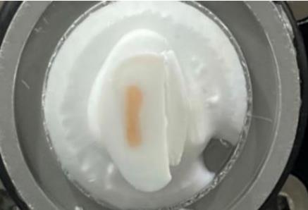
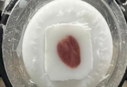
Embedding Tissue in OCT
After freezing, tissues are typically embedded in Optimal Cutting Temperature (OCT) compound, which provides structural support during cryosectioning.
Advantages of OCT Embedding
-
Stabilizes fragile tissues
Enables smooth cryosectioning
Preserves tissue morphology
Compatible with downstream staining and imaging
Embedding Tips
-
Ensure the tissue is fully surrounded by OCT
Remove air bubbles near the tissue
Maintain proper tissue orientation
Freeze the embedded block quickly
Accurate orientation is particularly important for spatial transcriptomics studies where anatomical context matters.
More detailed information on sample preparation protocols can be found here:BMKGENE Spatial Transcriptommics Sample Preparation Guidance
Shipping and Storage
Once samples are frozen:
-
Wrap blocks in aluminum foil
Seal them in a secure container
Maintain storage at −80 °C or in liquid nitrogen
For transportation, samples should be shipped on dry ice. Approximately 5 kg of dry ice per day of shipping is often recommended to maintain proper temperature during transit.
Common Issues to Avoid
Several common mistakes during preparation can compromise spatial transcriptomics experiments.
Delayed Freezing - Slow processing can result in RNA degradation and reduced data quality.
Incomplete OCT Embedding - If the tissue is not fully surrounded by OCT, the sample may break during cryosectioning.
Incorrect Tissue Orientation - Improper positioning may prevent the region of interest from appearing in sections.
Ice Crystal Formation - Improper freezing techniques can cause tissue distortion and poor morphology.
Final Thoughts
Successful spatial transcriptomics experiments begin with careful tissue handling and freezing. Rapid processing, proper OCT embedding, and correct freezing methods help preserve both RNA integrity and tissue architecture.
Both dry ice embedding and isopentane cooled with liquid nitrogen can produce high-quality results when performed correctly. Selecting the appropriate method depends on the tissue type, available equipment, and desired preservation quality.
Careful adherence to these preparation practices helps ensure that spatial transcriptomics experiments generate reliable and biologically meaningful gene expression maps.

Legal Disclaimer:
MENAFN provides the
information “as is” without warranty of any kind. We do not accept
any responsibility or liability for the accuracy, content, images,
videos, licenses, completeness, legality, or reliability of the information
contained in this article. If you have any complaints or copyright
issues related to this article, kindly contact the provider above.
Most popular stories
Market Research

More Story


Comments
No comment